RESEARCH ARTICLE
Siqi Wang · Jiayi Bai · Huiping Dai · Jie Zhan · Liping Ren · Brett H. Robinson · Chengzhi Jiang · Shuang Cui · Lidia Skuza · Shuhe Wei
Received: 30 November 2024 / Accepted: 24 March 2025 © The Author(s), under exclusive licence to Springer Nature Switzerland AG 2025
1Abstract摘要
Purpose We aimed to elucidate the speciation of Cd and Cu in the herbaceous flowering hyperaccumulator Bidens pilosa L. and their detoxification mechanisms. Methods Seedlings of B. pilosa were grown in hydroponic solution in a greenhouse. FTIR was used to determine differences in functional groups (including -OH, -CH2, -CH(CH3)2, -CO2H and -NH2) in molecular compounds from different tissue cells under different concentrations of Cd and Cu treatments. The subcellular distributions and chemical
目的 本研究旨在阐明草本开花超富集植物鬼针草(Bidens pilosa L.)体内Cd和Cu的形态及其解毒机制。方法 在温室中采用水培方式培育B. pilosa幼苗。利用FTIR测定不同浓度Cd和Cu处理下不同组织细胞中分子化合物官能团(包括-OH、-CH2、-CH(CH3)2、-CO2H和-NH2)的差异。采用差速离心法和连续提取法测定Cd和Cu的亚细胞分布及化学
Responsible Editor: Juan Barcelo.责任编辑:Juan Barcelo。
forms of Cd and Cu were determined by using differential centrifugation and sequential extraction methods.
形态。
Results FTIR revealed that the abundance of -OH, -CH2 groups, acid amides and lipids increased with increasing Cd concentrations. indicating that corresponding macromolecules, such as alcohols and phenols, proteins and lipids in cells in the stems/roots of B. pilosa reduced Cd stress. The results showed that -CH(CH3)2 groups bound Cd on the surface of the root cells. In contrast, the amount of -CH(CH3)2 groups was reduced with increasing Cu stress. The amount of -CH(CH3)2 groups and protein with acid
结果 FTIR结果表明,-OH、-CH2基团、酸酰胺和脂类的丰度随Cd浓度升高而增加,说明B. pilosa茎/根细胞中相应的大分子物质(如醇类和酚类、蛋白质及脂类)缓解了Cd胁迫。结果显示,-CH(CH3)2基团在根细胞表面结合Cd。相反,-CH(CH3)2基团的数量随Cu胁迫增强而减少。-CH(CH3)2基团及含酸
S. Wang (*) · C. Jiang · S. Cui Academy of Environmental and Chemical Engineering, Shenyang Ligong University, Shenyang 110159, Liaoning, China e-mail: wang3331336637@126.com
S. Wang · J. Bai · L. Ren · S. Wei (*)
S. Wang · J. Bai · L. Ren · S. Wei (*)
Key Laboratory of Pollution Ecology and Environment Engineering, Institute of Applied Ecology, Chinese Academy of Sciences, Shenyang 110016, China e-mail: shuhewei@iae.ac.cn
J. ZhanJ. Zhan
Liaoning Vocational College of Medicine, Shenyang 110101, China
辽宁医药职业学院,沈阳 110101,中国
B. H. RobinsonB. H. Robinson
School of Physical and Chemical Sciences, University of Canterbury, Christchurch 8041, New Zealand
坎特伯雷大学物理与化学科学学院,基督城 8041,新西兰
L. SkuzaL. Skuza
Institute of Biology, Centre for Molecular Biology and Biotechnology, University of Szczecin, 71-415 Szczecin, Poland
University of Szczecin生物研究所分子生物学与生物技术中心,71-415 Szczecin,波兰
H. Dai (*)H. Dai (*)
College of Biological Science & Engineering, Shaanxi Province Key Laboratory of Bio-Resources, Qinling- Bashan Mountains Bioresources Comprehensive Development C.I.C, State Key Laboratory of Biological Resources and Ecological Environment Jointly Built by Qinba Province and Ministry, Shaanxi University of Technology, Hanzhong 723001, China e-mail: daihp72@snut.edu.cn amides increased in cells in the stems of B. pilosa under Cd treatments. Subcellular distributions and chemical speciation analyses revealed that Cd was mainly sequestered in cytoplasm and cell walls of B. pilosa roots. In contrast, Cu was mostly bound in the cell walls. In the roots, the most Cd and Cu were ethanol extractable, indicating relative lability with a high potential to be translocated to the shoots. In the stems and leaves most Cd was bound in the cell walls. But there were no significant changes for the proportion of ethanol-extracted Cu in stems and leaves. Conclusions B. pilosa is a Cd hyperaccumulator, but not a Cu hyperaccumulator. The key compounds associated with Cd hyperaccumulation in B. pilosa were alcohols, phenols, acid amides and lipids.
陕西理工大学生物科学与工程学院,陕西省资源生物重点实验室,秦巴山区生物资源综合开发协同创新中心,省部共建秦巴生物资源与生态环境国家重点实验室,汉中 723001,中国 e-mail: daihp72@snut.edu.cn 酰胺的蛋白质数量在Cd处理下B. pilosa茎细胞中增加。亚细胞分布与化学形态分析表明,Cd主要被隔离于B. pilosa根的细胞质和细胞壁中;相反,Cu主要结合在细胞壁中。在根中,大部分Cd和Cu为乙醇提取态,表明其相对活跃,具有较高的向地上部转运潜力。在茎和叶中,大部分Cd结合于细胞壁中,而茎和叶中乙醇提取态Cu的比例无显著变化。结论 B. pilosa是Cd超富集植物,但不是Cu超富集植物。与B. pilosaCd超富集相关的关键化合物为醇类、酚类、酸酰胺和脂类。
Keywords Bidens pilosa L. · Cadmium · Copper · Functional groups · Subcellular distribution · Chemical form
关键词 Bidens pilosa L. · 镉 · 铜 · 官能团 · 亚细胞分布 · 化学形态
2Introduction引言
Hyperaccumulators and accumulators are plants, with heavy metal concentrations in the shoots that are several times higher than non-accumulator plants, without a significant reduction in biomass (Xu et al. 2022). Hyperaccumulator plants have mechanisms for both the translocation and tolerance of heavy metals. Plants that hyperaccumulate Cd and Cu are hypothesised to detoxify these elements through immobilisation on cell walls, chelation with organic acids from root exudates, sequestration in the the vacuole, the production of antioxidant enzymes, and increased synthesis of proteins, amino acids and polysaccharides (Heredia et al. 2022; Pasricha et al. 2021). Elucidating the mode and route of transport, and the mechanisms of Cd or Cu enrichment in accumulator and hyperaccumulator plants can inform the the management of contaminated sites (Feng et al. 2019; Yang et al. 2017). Yet there is a lacuna of information on the role of functional groups in accumulator and hyperaccumulator plants.
超富集植物和富集植物是指地上部重金属浓度比非富集植物高出数倍、而生物量无显著降低的植物(Xu et al. 2022)。超富集植物同时具备重金属转运和耐受机制。一般认为,超富集Cd和Cu的植物通过以下途径对这些元素进行解毒:细胞壁固定、根系分泌物中有机酸的螯合、液泡区隔化、抗氧化酶的产生,以及蛋白质、氨基酸和多糖合成的增加(Heredia et al. 2022; Pasricha et al. 2021)。阐明富集植物和超富集植物中Cd或Cu的转运方式与途径及富集机制,可为污染场地的管理提供依据(Feng et al. 2019; Yang et al. 2017)。然而,关于官能团在富集植物和超富集植物中作用的信息仍十分匮乏。
Black-jack (Bidens pilosa L.) is an annual species of herbaceous flowering plant in the asteraceae family. It is native to the tropical regions of America and widely distributed in tropical and subtropical regions of Asia. B. pilosa propagates by seeds that are transported by wind, water, and through mechanical adhesion to animals (Cai et al. 2022; Omara et al. 2020; Tolmacheva et al. 2014). Wei and Zhou (2008) reported that B. pilosa exhibited the characteristics of Cd hyperaccumulator accumulating and tolerating Cd concentrations manifold higher than other species growing in the same environment. Cd concentrations in B. pilosa leaves and stems were up to 192 mg/kg and 115 mg/kg, respectively, with soil Cd concentrations of 100 mg/kg. The authors also showed that B. pilosa was not an accumulator or hyperaccumulator of Cu, as shoot biomass decreased significantly compared to controls in soil containing 400 mg/kg Cu, while root and shoot Cu concentrations were 39 mg/kg and 12 mg/kg, respectively. Wei et al. (2018) showed that the root biomass and net photosynthetic rate increased significantly when B. pilosa was treated with 60 mg kg−1 Cd for five months, and Cd concentration in the shoots was up to 295 mg kg−1. Yu et al. (2022a) assessed the Cd uptake B. pilosa and five other plant species in a field study and found that the translocation factor (TF) of B. pilosa was significantly higher than those of the other plant species except Solanum nigrum L, reaching 2.46 ± 0.02. Sun et al. (2009) demonstrated that the growth of B. pilosa plants was promoted by soil Cd concentrations up to mg kg−1, with an increase in shoot biomass by 13.8% compared to the controls. The Cd concentrations of stems and leaves were 108 and 144 mg kg−1, respectively, at Cd soil concentration of 8 mg kg−1.
鬼针草(Bidens pilosa L.)为菊科一年生草本开花植物,原产于美洲热带地区,广泛分布于亚洲热带和亚热带地区。B. pilosa以种子繁殖,种子可借助风、水以及机械黏附于动物体表进行传播(Cai et al. 2022; Omara et al. 2020; Tolmacheva et al. 2014)。Wei and Zhou (2008)报道,B. pilosa表现出Cd超富集植物的特征,其对Cd的积累和耐受浓度比生长于同一环境中的其他物种高出数倍。在土壤Cd浓度为100 mg/kg时,B. pilosa叶和茎中的Cd浓度分别高达192 mg/kg和115 mg/kg。上述作者还表明,B. pilosa并非Cu富集植物或超富集植物,因为在含400 mg/kg Cu的土壤中,其地上部生物量较对照显著降低,而根和地上部Cu浓度分别为39 mg/kg和12 mg/kg。Wei et al. (2018)表明,B. pilosa经60 mg kg−1 Cd处理5个月后,根生物量和净光合速率显著增加,地上部Cd浓度高达295 mg kg−1。Yu et al. (2022a)在田间研究中评估了B. pilosa及其他5种植物对Cd的吸收,发现除Solanum nigrum L外,B. pilosa的转运系数(TF)显著高于其他植物,达2.46 ± 0.02。Sun et al. (2009)证实,土壤Cd浓度不超过 mg kg−1时可促进B. pilosa生长,地上部生物量较对照增加13.8%。在土壤Cd浓度为8 mg kg−1时,茎和叶的Cd浓度分别为108和144 mg kg−1。
Potentially, fourier transform infrared spectroscopy (FTIR) may be used to identify the composition of critical organic compounds that responsible Cd and Cu tolerance and accumulation (Yu et al. 2018, 2020; He et al. 2020). Using FTIR, Sharma and Uttam (2017) showed that the pectin content in the cell wall of wheat seedlings increased significantly under 500, 600, 700, and 800 mg L−1 nano-CuO stress, and the characteristic peaks at 1240 and 1320 cm−1 were altered with increasing nano-CuO concentrations. An increasing lignin content was determined in leaves on the basis of an increased band intensity at the area of 1507 cm−1. Reduced band areas of -CH2 lipid groups indicated that treatments with nano-CuO increased lipid peroxidation in plants. Liu et al. (2021) studied cell wall polysaccharides from beet (Beta vulgaris L.), kiwifruit (Actinidia chinensis Planch.), apple (Malus pumila Mill.) and found that the absorbance peaks at 1035 cm−1 were due to xylose-containing hemicelluloses, those at 1065 and 807 cm−1 were attributed to mannose-containing hemicelluloses, while the characteristic peaks at 1740 and 1600 cm−1 correspond to homogalacturonans based on the degree of methylation. Suresh et al. (2016) studied the effects of nano-CuO stress on biochemical constituents of peanut (Arachis hypogaea L.) leaves and found characteristic peaks at 1033, 1636 and 2923 cm−1, which corresponded to carbohydrates, proteins and lipids in leaf tissues, respectively. Sharma and Uttam (2019) studied the biochemical response of wheat (Triticum aestivum L.) seedling leaves to nano-Al2O3 stress and observed that the intensities of characteristic peaks corresponding to pectin, cellulose, lignin and hemicellulose were significantly more intense as a result of 3 mM nano-Al2O3 treatments, and the peak areas related to methylene and amine groups visibly increased with raising nano-Al2O3 concentrations, which indicated that the defense strategy of wheat seedlings against nano-Al2O3 toxicity was based on improving lipids, amino acids and proteins in leaves. Yu et al. (2020) characterised the physiological responses of Conyza canadensis (L.) Cronq. to 1 ~ 7 mg L−1 of Cd stress using FTIR and found three characteristic peaks (3417 ~ 3429 cm−1, 1380 ~ 1386 cm−1, and 1631 ~ 1637 cm−1), which corresponded to hydroxy, carboxyl and acid amide groups. These functional groups are all involved in Cd binding and adsorption. Chow and Ting (2019) found that changes in the plant cell wall chemical composition could be inferred using FTIR and the intensity ratios of absorption peaks (I1458/I1158 and I1458/I1375), which corresponded to the synthesis and decomposition of lignin and carbohydrates. Yu et al. (2021) demonstrated that the removal of pectins markedly reduced the peak areas at 3477 cm−1, 3414 and 1619 cm−1 in the cell wall of rice (Oryza sativa L.) roots, which corresponded to the lower amount of carboxyl, hydroxyl, and nitrogen–hydrogen bonds of amines, and carbonyl amide I functional groups. The latter results suggested that the removal of hemicellulose and pectin decreased the relative peak areas at 1251, 1430, 1515, 1730 and 2920 cm−1. These authors also investigated the mechanisms of Cd binding to polysaccharides in the root cell wall of Cdexcluding rice cultivars using FTIR and suggested that the carboxyl and hydroxyl groups in pectin and hemicellulose were beneficial for Cd immobilization and retention.
傅里叶变换红外光谱(FTIR)有望用于鉴定与Cd和Cu耐受及积累相关的关键有机化合物组成(Yu et al. 2018, 2020; He et al. 2020)。Sharma and Uttam (2017)利用FTIR研究发现,在500、600、700和800 mg L−1纳米CuO胁迫下,小麦幼苗细胞壁中果胶含量显著增加,且1240和1320 cm−1处的特征峰随纳米CuO浓度升高而发生改变。根据1507 cm−1区域谱带强度的增加,测定出叶片中木质素含量升高。-CH2脂类基团谱带面积的减小表明,纳米CuO处理加剧了植物的脂质过氧化。Liu et al. (2021)研究了甜菜(Beta vulgaris L.)、猕猴桃(Actinidia chinensis Planch.)和苹果(Malus pumila Mill.)的细胞壁多糖,发现1035 cm−1处的吸收峰源于含木糖的半纤维素,1065和807 cm−1处的吸收峰归属于含甘露糖的半纤维素,而1740和1600 cm−1处的特征峰则根据甲基化程度对应于同型半乳糖醛酸聚糖。Suresh et al. (2016)研究了纳米CuO胁迫对花生(Arachis hypogaea L.)叶片生化成分的影响,发现1033、1636和2923 cm−1处的特征峰分别对应叶组织中的碳水化合物、蛋白质和脂类。Sharma and Uttam (2019)研究了小麦(Triticum aestivum L.)幼苗叶片对纳米Al2O3胁迫的生化响应,观察到3 mM纳米Al2O3处理使果胶、纤维素、木质素和半纤维素对应特征峰的强度显著增强,且亚甲基和胺基相关峰面积随纳米Al2O3浓度升高而明显增加,表明小麦幼苗抵御纳米Al2O3毒性的防御策略在于提高叶片中的脂类、氨基酸和蛋白质。Yu et al. (2020)利用FTIR表征了Conyza canadensis (L.) Cronq.对1 ~ 7 mg L−1 Cd胁迫的生理响应,发现3个特征峰(3417 ~ 3429 cm−1、1380 ~ 1386 cm−1和1631 ~ 1637 cm−1),分别对应羟基、羧基和酸酰胺基团,这些官能团均参与Cd的结合与吸附。Chow and Ting (2019)发现,利用FTIR及吸收峰强度比(I1458/I1158和I1458/I1375),其对应于木质素和碳水化合物的合成与分解)可推断植物细胞壁化学组成的变化。Yu et al. (2021)证实,去除果胶显著降低了水稻(Oryza sativa L.)根细胞壁中3477 cm−1、3414和1619 cm−1处的峰面积,这对应于羧基、羟基、胺的氮氢键以及羰基酰胺I官能团数量的减少。后一结果表明,去除半纤维素和果胶降低了1251、1430、1515、1730和2920 cm−1处的相对峰面积。上述作者还利用FTIR研究了Cd排斥型水稻品种根细胞壁多糖结合Cd的机制,认为果胶和半纤维素中的羧基和羟基有利于Cd的固定与滞留。
Zou et al. (2023) reported that Ca addition could alleviate physiological toxicity of Chinese willow trees (Salix matsudana Koidz) under Cd stress, decrease Cd uptake by roots, increase transport from root to shoot, elevate the proportion of Cd in the cytoplasm and reduce Cd deposition in cell wall, and lead to Cd subcellular redistribution. The chemical form of Cd was changed from HCl-extracted to NaCl-extracted. Li et al (2023) investigated the subcellular mechanism of Cd accumulation by black nightshade (S. nigrum L.), and found that vacuolar compartmentalisation was a dynamic regulatory process with continuous glutathione consumption, and the role of glutathione in vacuoles was to chelate and store Cd. Wu et al. (2016) investigated the principles of Cd accumulation, localization, subcellular distribution and chemical forms in Salix matsudana Koid roots, and found that Cd concentration in the different subcellular fractions was in the following order: cell wall > cytoplasm > organelle, after treatment with 100 μM Cd for 24 h. Meanwhile, the proportion of Cd chemical forms in roots was ordered as follows: NaCl-extracted > CH3COOH-extracted > HCl-extracted > water-extracted > ethanol-extracted > residue-extracted. Teng et al. (2021) studied the detoxification mechanism and vacuolar compartmentalizationin in the leaf of S. nigrum, and discovered that the majority of dissolved Cd ions or combined Cd was trapped inside or outside of the vacuoles. More than 99% Cd was stored in vacuoles after treatment with 25 μM Cd. Furthermore, and 3 ~ 10 times of Cd accumulation was combined inside the vacuole than that of outside.
Zou et al. (2023)报道,添加Ca可缓解Cd胁迫下旱柳(Salix matsudana Koidz)的生理毒性,降低根系对Cd的吸收,增加由根向地上部的转运,提高细胞质中Cd的比例并减少Cd在细胞壁中的沉积,从而导致Cd的亚细胞再分配;Cd的化学形态由HCl提取态转变为NaCl提取态。Li et al (2023)研究了龙葵(S. nigrum L.)积累Cd的亚细胞机制,发现液泡区隔化是一个伴随谷胱甘肽持续消耗的动态调控过程,液泡中谷胱甘肽的作用是螯合并储存Cd。Wu et al. (2016)研究了Salix matsudana Koid根中Cd的积累、定位、亚细胞分布及化学形态规律,发现经100 μM Cd处理24 h后,不同亚细胞组分中的Cd浓度依次为:细胞壁 > 细胞质 > 细胞器;同时,根中各化学形态Cd的比例依次为:NaCl提取态 > CH3COOH提取态 > HCl提取态 > 水提取态 > 乙醇提取态 > 残渣态。Teng et al. (2021)研究了S. nigrum叶片中的解毒机制与液泡区隔化,发现大部分溶解态Cd离子或结合态Cd被截留于液泡内外。经25 μM Cd处理后,超过99%的Cd储存于液泡中;此外,液泡内结合的Cd积累量为液泡外的3 ~ 10倍。
Some studies have focused on Cd accumulation by B. pilosa. However, there are no reports on the differences between Cu and Cd detoxification by B. pilosa. Therefore, the aims of this study were: (1) to determine the differences between Cd hyperaccumulation by B. pilosa compared with Cu; (2) to identify compounds and functional groups of B. pilosa associated with elevated Cd in relation to Cu by FTIR, with special attention to differences in the functional group characteristics; (3) to understand the mechanism behind Cd and Cu resistance in B. pilosa by investigating the subcellular distribution and chemical forms. It was hypothesized that the functional groups characteristics, subcellular distributions and chemical forms would show significant differences in the mechanisms of B. pilosa Cd hyperaccumulation and detoxification compared to Cu.
已有一些研究关注B. pilosa对Cd的积累,但尚无关于B. pilosa对Cu和Cd解毒差异的报道。因此,本研究的目的是:(1)确定B. pilosa对Cd超富集与对Cu富集的差异;(2)利用FTIR鉴定B. pilosa中与高Cd(相对于Cu)相关的化合物和官能团,尤其关注官能团特征的差异;(3)通过研究亚细胞分布和化学形态,阐明B. pilosa抗Cd和抗Cu的机制。本研究假设:与Cu相比,B. pilosa在Cd超富集和解毒机制上,其官能团特征、亚细胞分布和化学形态均存在显著差异。
3Materials and methods材料与方法
Plant culture and treatment植物培养与处理
A series of experiments was carried out in a greenhouse located in the Shenyang Institute of Applied Ecology of the Chinese Academy of Sciences (123°59′E and 41°92′N). B. pilosa seeds were collected from the surrounding park, which has uncontaminated soil, when they reached the maturity phase. On July 4th, 2024, twenty seeds of B. pilosa were sown after surface disinfection in nursery pots (20 cm × 30 cm × 5 cm) filled with sterile sand (quartz sand, AR, 14808–60-7, particle size was 0.3 mm ~ 0.6 mm, National Pharmaceutical Group Corporation, Shanghai, China) to a depth of 2 cm. Seeds were cultivated in a greenhouse with a constant temperature (24 ± 2℃) at an 8/16 h dark/light photoperiod. Half strength Hoagland solution (100 mL) was added every two days. When the height of most seedlings reached 5 cm, uniform seedlings were transplanted (11 per container) to transparent plastic containers (25 cm × 10 cm × 16 cm) to observe root growth; containers were filled with sterile 1/2-strength Hoagland solution with Cd or Cu under 24 h aeration with an ACO-9610 adjustable silent oxygen pump (Hailea, Guangdong, China), and 0.025 mM KH2PO4 was replaced with KCl to avoid the formation of Cd or Cu phosphate precipitates in the solution. The roots of B. pilosa seedlings were stressed with 0.5, 2, 8, 16 mg L−1 Cd (as CdCl2 ‧ 2.5 H2O) and Cu (as CuSO4 ‧ 5 H2O) treatments. The pH of the solutions was maintained at 5.5 ± 0.2 by adding 2 mM MES (2-morpholinoethanesulphonic acid). Sterile nutrient solutions without Cd or Cu were used as controls, and the solutions from each container were replaced every three days. Each treatment was repeated thrice and containers were arranged in a randomized block design in the experiments. Twenty-five containers were rotated every 12 h to maintain uniform environmental conditions. The plants were harvested seven days after transplanting. Meanwhile, every repetition of treatments was analyzed three times from July 15th to August 15th, 2024.
系列试验在中国科学院沈阳应用生态研究所温室(123°59′E,41°92′N)内进行。B. pilosa种子于成熟期采自周边土壤未受污染的公园。2024年7月4日,将20粒经表面消毒的B. pilosa种子播于装有无菌砂(石英砂,AR,14808–60-7,粒径0.3 mm ~ 0.6 mm,国药集团化学试剂有限公司,上海,中国)的育苗盆(20 cm × 30 cm × 5 cm)中,播种深度为2 cm。种子在恒温(24 ± 2℃)、暗/光周期为8/16 h的温室中培养,每两天添加半强度Hoagland营养液(100 mL)。当大多数幼苗高度达到5 cm时,将长势一致的幼苗移栽(每个容器11株)至透明塑料容器(25 cm × 10 cm × 16 cm)中以便观察根系生长;容器中装有含Cd或Cu的无菌1/2强度Hoagland营养液,使用ACO-9610可调静音增氧泵(Hailea,广东,中国)24 h通气,并以KCl替代0.025 mM KH2PO4,以避免溶液中形成Cd或Cu的磷酸盐沉淀。B. pilosa幼苗根系分别接受0.5、2、8、16 mg L−1 Cd(以CdCl2 ‧ 2.5 H2O形式)和Cu(以CuSO4 ‧ 5 H2O形式)胁迫处理。通过添加2 mM MES(2-吗啉乙磺酸)将溶液pH维持在5.5 ± 0.2。以不含Cd或Cu的无菌营养液为对照,每个容器中的溶液每三天更换一次。每个处理重复3次,容器按随机区组设计排列。25个容器每12 h轮换一次位置,以保持环境条件一致。移栽7天后收获植株。同时,于2024年7月15日至8月15日期间,对各处理的每个重复分析3次。
FTIR spectra and determination of biomass and Cd/ Cu concentration
FTIR光谱及生物量和Cd/ Cu浓度的测定
Briefly, roots, stems and leaves of B. pilosa seedlings were separated, freeze-dried, pulverized with KBr (v/v = 1:100) and sieved through a 200-µm mesh, FTIR spectral data (4000 to 400 cm−1) of individual tissue parts were obtained using a FTIR spectrometer (Nicolet 6700, Thermo Fisher Scientific, Massachusetts, USA).
简言之,将B. pilosa幼苗的根、茎和叶分离,冷冻干燥后与KBr(v/v = 1:100)混合研磨并过200-µm筛,使用FTIR光谱仪(Nicolet 6700,Thermo Fisher Scientific,Massachusetts,USA)获取各组织部位的FTIR光谱数据(4000至400 cm−1)。
The collected roots and shoots of B. pilosa were separated and washed with ultrapure water. The samples were oven-dried at 100℃ for 30 min, and then at 80℃ to a constant weight. The biomass was measured using a balance to the nearest 0.001 g The samples of roots, stems and leaves subjected to various treatments were digested according to a method described by Zeng et al. (2019), with modifications; Cd and Cu concentrations were determined using ICP-OES (iCAP™ 7200, Thermo Fisher Scientific, Massachusetts, USA) (Hamid et al. 2019). The measured Cd concentration in B. pilosa seedlings was checked based on the standard reference material for plant composition analysis (GBW07604, GSV-3, poplar leaves) (Wei et al. 2010). The blanks and samples were determined in triplicate. The limits of detection (LOD) and quantification (LOQ) were 0.0018 and 0.0037 mg/kg, respectively. The measured values of Cd and Cu in certified materials were 0.29 ± 0.01 and 9.3 ± 1.0 mg/kg, and the values of relative standard deviation (RSD) and Cd recovery rate were 3% and 90 ± 1%, respectively.
将收获的B. pilosa根和地上部分离,用超纯水洗净。样品先于100℃烘箱中杀青30 min,再于80℃烘至恒重,用精度为0.001 g的天平测定生物量。各处理的根、茎、叶样品参照Zeng et al. (2019)所述方法(略有修改)进行消解;采用ICP-OES(iCAP™ 7200,Thermo Fisher Scientific,Massachusetts,USA)测定Cd和Cu浓度(Hamid et al. 2019)。以植物成分分析标准物质(GBW07604,GSV-3,杨树叶)对B. pilosa幼苗中Cd浓度的测定结果进行质量控制(Wei et al. 2010)。空白和样品均测定3次。检出限(LOD)和定量限(LOQ)分别为0.0018和0.0037 mg/kg。标准物质中Cd和Cu的测定值分别为0.29 ± 0.01和9.3 ± 1.0 mg/kg,相对标准偏差(RSD)和Cd回收率分别为3%和90 ± 1%。
The translocation factor (TF) was determined on the basis of the ratio of the Cd shoot to root Cd concentration according to the following formula (2.1): TF = Cdshoot (2.1) Cdroot
转运系数(TF)根据地上部Cd浓度与根部Cd浓度之比,按下式(2.1)计算:TF = Cdshoot (2.1) Cdroot
Analysis of subcellular distribution of Cd and Cu
Cd和Cu的亚细胞分布分析
The subcellular tissues of roots, stems and leaves from B. pilosa seedlings were separated by differential centrifugation methods (Pan et al. 2019; Luo et al. 2024). Firstly, 0.5 g fresh samples were frozen with liquid nitrogen, and ground with a chilled mortar and pestle, and then homogenized with 5 mL of 50 mM Tris–HCl (pH = 7.5) and 1 mM C₄HO₂S₂ (DTT) as extraction buffers. Secondly, after the samples were centrifuged at 600 × g for 10 min, the cell wall (FI) was obtained from the precipitate. The supernatants were further centrifuged at 2000 × g for 10 min, and the resulting precipitate representing the plastid (chloroplasts in leaves) (FII). Thirdly, the remaining supernatants were further centrifuged at 10,000 × g for 20 min, and the resulting precipitates and supernatants representing the mitochondria (FIII) and cytoplasm (FIV), respectively. The above centrifugation procedures were all completed using high speed centrifuge (Microfuge 20/20R, Beckman Coulter, Brea, California, USA) at 4 ℃. Meanwhile, the Cd/Cu concentrations in three fractions were determined according to the method used to analyze plant Cd/Cu concentrations.
采用差速离心法分离B. pilosa幼苗根、茎和叶的亚细胞组分(Pan et al. 2019; Luo et al. 2024)。首先,取0.5 g新鲜样品经液氮冷冻后,用预冷的研钵和研杵研磨,再以5 mL含50 mM Tris–HCl(pH = 7.5)和1 mM C₄HO₂S₂(DTT)的提取缓冲液匀浆。其次,样品在600 × g下离心10 min,沉淀为细胞壁组分(FI)。上清液再于2000 × g下离心10 min,所得沉淀为质体组分(叶片中为叶绿体)(FII)。第三,剩余上清液于10,000 × g下离心20 min,所得沉淀和上清液分别为线粒体组分(FIII)和细胞质组分(FIV)。上述离心步骤均在4 ℃下使用高速离心机(Microfuge 20/20R,Beckman Coulter,Brea,California,USA)完成。同时,按照植物Cd/Cu浓度的分析方法测定各组分中的Cd/Cu浓度。
Extraction of Cd and Cu in different chemical forms
不同化学形态Cd和Cu的提取
Different Cd and Cu chemical forms of roots, stems and leaves from B. pilosa seedlings were extracted by sequential extraction method (Pan et al. 2019; An et al. 2023). 0.5 g fresh samples were frozen and ground, and then homogenized with different extraction solutions (20 mL) as follows: (1) 80% ethanol; (2) deionized water; (3) 1 M NaCl; (4) 2% CH3COOH; (5) 0.6 M HCl; (6) Residues. Firstly, the different homogenates were shaken for 24 h at 24 ºC and centrifuged at 5000 rpm for 10 min, the first supernatant was collected in a conical beaker. Meanwhile, 10 mL corresponding extraction solution was added into the precipitate was shaken for 2 h at 24 ºC. Secondly, the homogenate was centrifuged at 5000 rpm for 10 min, the second supernatant was mixed with the first one as tested solution, and the second precipitates were taken as residues. Finally, different chemical forms of Cd and Cu were obtained by the above procedures. The Cd/Cu concentrations in second precipitates were determined according to the method used to analyze plant Cd/Cu concentrations.
采用连续提取法提取B. pilosa幼苗根、茎和叶中不同化学形态的Cd和Cu(Pan et al. 2019; An et al. 2023)。取0.5 g新鲜样品冷冻研磨后,依次用以下提取液(20 mL)匀浆:(1)80%乙醇;(2)去离子水;(3)1 M NaCl;(4)2% CH3COOH;(5)0.6 M HCl;(6)残渣。首先,各匀浆液在24 ºC下振荡24 h,于5000 rpm离心10 min,将第一次上清液收集于锥形烧杯中;同时,向沉淀中加入10 mL相应提取液,在24 ºC下振荡2 h。其次,匀浆液于5000 rpm离心10 min,将第二次上清液与第一次上清液合并作为待测液,第二次沉淀作为残渣。最后,通过上述步骤获得不同化学形态的Cd和Cu。按照植物Cd/Cu浓度的分析方法测定第二次沉淀中的Cd/Cu浓度。
Data processing and statistical analysis数据处理与统计分析
Microsoft Excel 2022 and SPSS (26.0) software was used to calculate the means and standard deviations of the data (n = 3). LSD was used to test the inconsistencies in FTIR results (e.g., peak intensity, peak shift) under different Cd and Cu treatments, and the significant differences (p < 0.05) for individual treatment were assessed by using DPS (V9.50) (Pan et al. 2016; Wang et al. 2022). The determination and quantitative analysis of characteristic peaks in FTIR spectra of B. pilosa was conducted using Origin (V10.50).
采用Microsoft Excel 2022和SPSS(26.0)软件计算数据的平均值和标准差(n = 3)。采用LSD检验不同Cd和Cu处理下FTIR结果(如峰强度、峰位移)的差异,并使用DPS(V9.50)评估各处理间的显著性差异(p < 0.05)(Pan et al. 2016; Wang et al. 2022)。B. pilosa FTIR光谱特征峰的测定与定量分析使用Origin(V10.50)完成。
4Results结果
B. pilosa hyperaccumulation characteristics for CdB. pilosa对Cd的超富集特征
Table 1 Biomass and Cd and Cu concentration of Bidens pilosa L表1 Bidens pilosa L的生物量及Cd和Cu浓度
| Biomass (g p生物量 (g p | ot−1) | Concentration (mg kg−浓度 (mg kg− | 1) | TF | |
|---|---|---|---|---|---|
| Treatment | Root | Shoot | Root | Shoot | |
| Control | 0.94 ± 0.07a | 0.84 ± 0.04a | 2.92 ± 0.78e | 3.56 ± 0.18e | 1.28 ± 0.36 |
| Cd 0.5 | 0.90 ± 0.07a | 0.81 ± 0.03a | 78.82 ± 4.09d | 99.64 ± 5.42d | 1.26 ± 0.02 |
| Cd 2 | 0.91 ± 0.08a | 0.79 ± 0.01a | 483.42 ± 7.16c | 848.47 ± 169.66c | 1.76 ± 0.37 |
| Cd 8 | 0.71 ± 0.04b | 0.55 ± 0.03b | 4797.66 ± 54.96b | 5100.69 ± 284.48b | 1.06 ± 0.05 |
| Cd 16 | 0.65 ± 0.04c | 0.43 ± 0.03c | 6236.95 ± 71.44a | 7764.55 ± 441.39a | 1.24 ± 0.06 |
| Control | 0.94 ± 0.07a | 0.84 ± 0.04a | 12.31 ± 1.16c | 11.86 ± 1.16b | 0.97 ± 0.09 |
| Cu 0.5 | 0.76 ± 0.08b | 0.65 ± 0.07b | 55.60 ± 22.73c | 18.28 ± 0.95b | 0.37 ± 0.17 |
| Cu 2 | 0.36 ± 0.02c | 0.54 ± 0.08bc | 1477.65 ± 88.05c | 28.75 ± 1.86b | 0.02 ± 0.01 |
| Cu 8 | 0.29 ± 0.02c | 0.49 ± 0.07 cd | 9478.60 ± 1484.25b | 154.47 ± 44.01a | 0.02 ± 0.01 |
| Cu 16 | 0.17 ± 0.02d | 0.41 ± 0.06d | 12,322.18 ± 1929.53a | 213.66 ± 60.90a | 0.02 ± 0.01 |
Means of columns in different Cd or Cu treatments and controls marked with the same lowercase letters were not significantly different at p < 0.05 (from 0.5 to 16 mg L−1), and root and shoot biomass was reduced by 82% and 51% in 16 mg L−1 Cu treatments compared to controls.
不同Cd或Cu处理及对照中标有相同小写字母的各列平均值在p < 0.05水平上无显著差异(0.5至16 mg L−1),在16 mg L−1 Cu处理中,根和地上部生物量较对照分别降低82%和51%。
Cd accumulation in B. pilosa roots and shoots increased with increasing Cd concentrations relative to controls (Table 1). Cd concentrations in shoots were significantly higher than in roots after individual Cd treatments. The TF of B. pilosa was always > 1 for all Cd treatments, indicating its compliance with basic characteristics of a Cd hyperaccumulator. The highest TF was recorded in the treatments with 2 mg L−1 Cd. Cu accumulation in B. pilosa roots and shoots raised with increasing Cu concentrations in comparison to controls. Cu concentrations in shoots were significantly lower than in roots in individual Cu treatments. The TF was always < 1 in all Cu treatments, indicating that this plant was not a Cu hyperaccumulator.
与对照相比,B. pilosa根和地上部的Cd积累量随Cd浓度升高而增加(表1)。各Cd处理下,地上部Cd浓度均显著高于根部。所有Cd处理中B. pilosa的TF均 > 1,表明其符合Cd超富集植物的基本特征。TF最高值出现在2 mg L−1 Cd处理中。与对照相比,B. pilosa根和地上部的Cu积累量随Cu浓度升高而增加。各Cu处理下,地上部Cu浓度均显著低于根部。所有Cu处理中TF均 < 1,表明该植物不是Cu超富集植物。
FTIR differences between B. pilosa accumulating Cd and Cu
B. pilosa积累Cd与Cu的FTIR差异
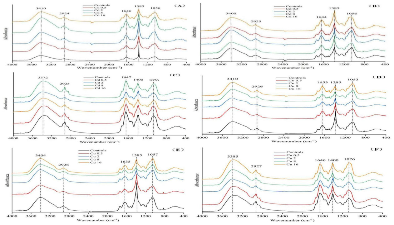
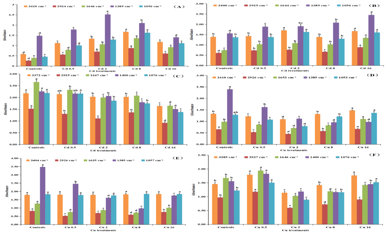
Functional groups in individual parts of B. pilosa under different Cd concentrations are shown in Fig. 1A, B, C. We selected five relatively significant characteristic peaks (3394 ± 20 cm−1, 2925 ± 1 cm−1, 1646 ± 2 cm−1, 1390 ± 9 cm−1 and 1063 ± 12 cm−1) from the range between 4000 and 400 cm−1, which corresponded to the following functional groups and biochemical regions: alcohols and phenols, composed of -OH (3700 ~ 3200 cm−1) and -CH2 groups (anti-symmetric C-H stretching vibration bands at 2930 cm−1), protein with acid amides (1700 ~ 1600 cm−1), -CH(CH3)2 groups (symmetric C-H stretching vibration bands at 1380 cm−1), and lipids, consisting of fatty acid long chains (C-O stretching vibration bands at 1300 ~ 1000 cm−1) (Yu et al. 2018, 2020, 2022b). As shown in Fig. 2A, the peaks at 3410, 2924, 1646 and 1056 cm−1 in the root spectrum reached their maximum in 8 mg L−1 Cd treatment, the abundance of -OH, -CH2 groups, acid amides and lipids increased with increasing Cd concentrations, indicating their involvement in the root cell co-precipitation and complexation in response to Cd toxicity. There was also a significant increase in 1385 cm−1 peaks at a Cd concentration of 2 mg L−1, indicating that the amount of -CH(CH3)2 groups was relatively higher compared to controls and other Cd treatments. The absorbance intensities of peaks at 2925 cm−1, 1644 and 1385 cm−1 of the stem spectrum gradually raised with increasing Cd stress concentrations. The maximum occurred at Cd concentration of 16 mg L−1; peak intensities at 3400 and 1056 cm−1 under 2 mg L−1 Cd stress were also clearly increased compared to other Cd treatments (Fig. 2B). The absorbance intensities of five characteristic peaks in the leaf spectrum decreased significantly in the 16 mg L−1 Cd treatments (Fig. 2C).
不同Cd浓度下B. pilosa各部位的官能团见图1A、B、C。我们在4000至400 cm−1范围内选取了5个相对显著的特征峰(3394 ± 20 cm−1、2925 ± 1 cm−1、1646 ± 2 cm−1、1390 ± 9 cm−1和1063 ± 12 cm−1),分别对应以下官能团和生化区域:由-OH(3700 ~ 3200 cm−1)组成的醇类和酚类,-CH2基团(2930 cm−1处的C-H反对称伸缩振动带),含酸酰胺的蛋白质(1700 ~ 1600 cm−1),-CH(CH3)2基团(1380 cm−1处的C-H对称伸缩振动带),以及由脂肪酸长链构成的脂类(1300 ~ 1000 cm−1处的C-O伸缩振动带)(Yu et al. 2018, 2020, 2022b)。如图2A所示,根光谱中3410、2924、1646和1056 cm−1处的峰在8 mg L−1 Cd处理中达到最大值,-OH、-CH2基团、酸酰胺和脂类的丰度随Cd浓度升高而增加,表明它们参与了根细胞响应Cd毒性的共沉淀和络合作用。在Cd浓度为2 mg L−1时,1385 cm−1处的峰也显著增强,表明-CH(CH3)2基团数量较对照及其他Cd处理相对较高。茎光谱中2925 cm−1、1644和1385 cm−1处峰的吸收强度随Cd胁迫浓度升高而逐渐增加,最大值出现在Cd浓度为16 mg L−1时;在2 mg L−1 Cd胁迫下,3400和1056 cm−1处的峰强度也较其他Cd处理明显增加(图2B)。在16 mg L−1 Cd处理中,叶光谱中5个特征峰的吸收强度显著降低(图2C)。
Bidens pilosa L. seedlings and controls. (Means in different treatments marked with the same lowercase letters are not significantly different at p < 0.05)(Bidens pilosa L.)。(不同处理中标有相同小写字母的平均值在p < 0.05水平上无显著差异)
Absorbance spectra of the roots, stems and leaves of B. pilosa were determined and analyzed in response to different Cu levels (Fig. 1D, E, F). The characteristic peaks could be observed by analyzing the infrared spectra (He et al. 2020; Mwamba et al. 2016). The absorbance peaks at 3394 ± 20 cm−1 belonged to the stretching vibration of -OH groups, dominant in phenols and alcohols, which were attributed to macromolecular substances, such as cellulose and hemicellulose polysaccharides. The characteristic peaks at 2925 ± 1 cm−1 were signals representing -CH2 groups, which could be caused by the presence of membrane lipids and a cell wall containing a large number of lipid compounds. The characteristic peaks at 1645 ± 9 cm−1 were acid amides present in the protein conformation. The absorbance peaks at 1390 ± 9 cm−1 were attributed to -CH(CH3)2 groups, and the peaks near 1062 ± 12 cm−1 represented C-O stretching vibrations commonly found in long-chain fatty acids, mainly from polyols present in carbohydrates from the cell wall. Compared to controls, the shapes, trends and positions of the peaks in the spectra of B. pilosa root, stem and leaf tissues were generally consistent, but several significant differences in peak intensities were still observed between individual Cu treatments. Figure 2D shows that the maximum absorbance intensities of the peaks at 3410 and 1653 cm−1 of the root spectrum occurred at Cu concentration of 16 mg L−1. The peak intensities at 1385 cm−1 gradually decreased with increasing Cu stress, and there were no significant differences in the peaks at 1053 and 2926 cm−1 under 16 mg L−1 Cd compared to controls. As shown in Fig. 2E, the intensities of the peaks at 2926, 1635 and 1385 cm−1 (-CH2, acid amides, -CH(CH3)2 groups) of the stem spectrum decreased significantly compared with controls, and there were no significant variations between the peaks at 3404 and 1057 cm−1 compared to controls. Figure 2F indicates that the peak intensities at 2927 and 1646 cm−1 of the leaf spectrum were lower in plants subject to Cu stress in the range of 2 ~ 16 mg L−1 than in controls, and there were no significant variations between the peaks at 1400 cm−1 under 16 mg L−1 Cu compared to controls. The maximum intensities of the peaks at 3385 and 1076 cm−1 were recorded at Cu concentrations of 0.5 or 16 mg L−1.
测定并分析了B. pilosa根、茎和叶在不同Cu水平下的吸收光谱(图1D、E、F)。通过分析红外光谱可观察到各特征峰(He et al. 2020; Mwamba et al. 2016)。3394 ± 20 cm−1处的吸收峰属于-OH基团的伸缩振动,主要存在于酚类和醇类中,归属于纤维素和半纤维素多糖等大分子物质。2925 ± 1 cm−1处的特征峰代表-CH2基团信号,可能源于膜脂以及含有大量脂类化合物的细胞壁。1645 ± 9 cm−1处的特征峰为蛋白质构象中的酸酰胺。1390 ± 9 cm−1处的吸收峰归属于-CH(CH3)2基团,1062 ± 12 cm−1附近的峰代表长链脂肪酸中常见的C-O伸缩振动,主要来自细胞壁碳水化合物中的多元醇。与对照相比,B. pilosa根、茎和叶组织光谱中峰的形状、趋势和位置总体一致,但各Cu处理间峰强度仍存在若干显著差异。图2D显示,根光谱中3410和1653 cm−1处峰的最大吸收强度出现在Cu浓度为16 mg L−1时。1385 cm−1处的峰强度随Cu胁迫增强而逐渐降低,而在16 mg L−1 Cd下,1053和2926 cm−1处的峰与对照相比无显著差异。如图2E所示,茎光谱中2926、1635和1385 cm−1处峰(-CH2、酸酰胺、-CH(CH3)2基团)的强度较对照显著降低,而3404和1057 cm−1处的峰与对照相比无显著变化。图2F表明,在2 ~ 16 mg L−1 Cu胁迫范围内,叶光谱中2927和1646 cm−1处的峰强度低于对照,而在16 mg L−1 Cu下,1400 cm−1处的峰与对照相比无显著变化。3385和1076 cm−1处峰的最大强度出现在Cu浓度为0.5或16 mg L−1时。
In the absorbance spectra of B. pilosa root, the peak intensity at 1385 cm−1 gradually increased with increasing Cd stress, but decreased under Cu stress. The results indicated that a number of -CH(CH3)2 groups were produced to combine with Cd in root cell surface. In contrast, the amount of -CH(CH3)2 groups was reduced with increasing Cu stress. In the same way, the amount of -CH(CH3)2 groups and protein with acid amides (at 1644 cm−1 and 1635 cm−1) were increased in B. pilosa cells in the stems of B. pilosa in the Cd treatments.
在B. pilosa根的吸收光谱中,1385 cm−1处的峰强度随Cd胁迫增强而逐渐升高,但在Cu胁迫下降低。结果表明,根细胞表面产生了大量-CH(CH3)2基团与Cd结合;相反,-CH(CH3)2基团的数量随Cu胁迫增强而减少。同样,在Cd处理中,B. pilosa茎中B. pilosa细胞内的-CH(CH3)2基团及含酸酰胺的蛋白质(1644 cm−1和1635 cm−1处)数量增加。
Subcellular distribution and different chemical forms of Cd and Cu in B. pilosa seedling
B. pilosa幼苗中Cd和Cu的亚细胞分布及不同化学形态
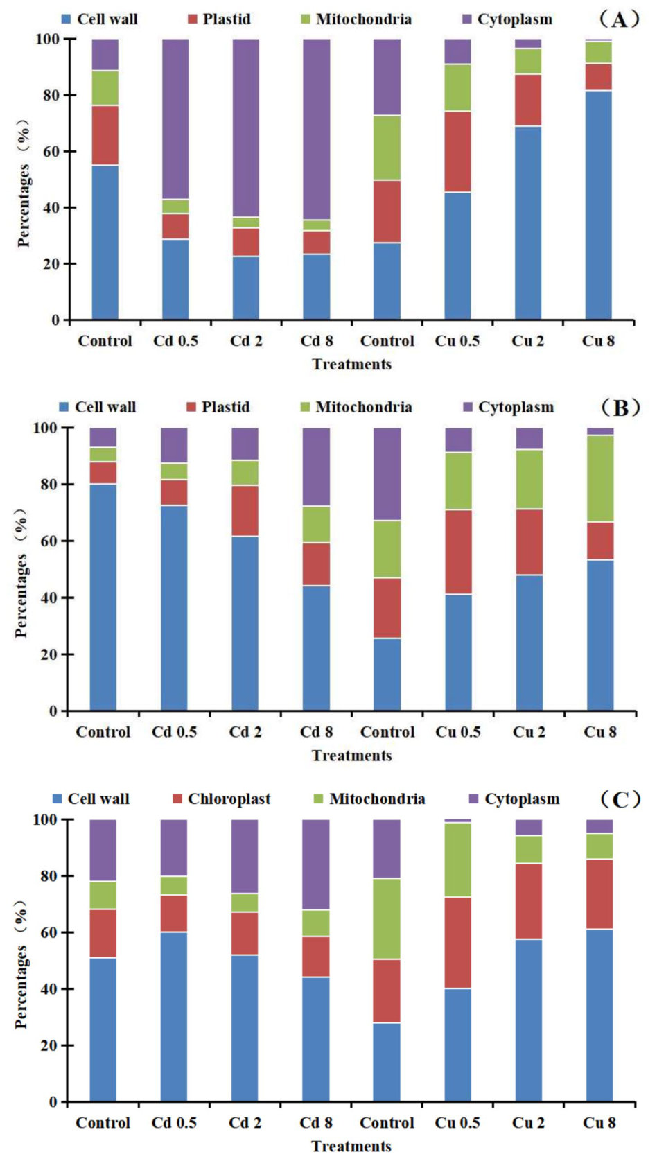
The percentages of Cd/Cu concentrations in different subcellular fractions from root, stem, and leaf tissues of B. pilosa seedlings under different treatments were shown from Fig. 3, and the order of Cd proportion in subcellular fractions from roots was: cytoplasm (61.73%) > cell wall (24.93%) > plastid (9.15%) > mitochondria (4.19%) (Fig. 3A). The Cd proportion in cell wall decreased, while in cytoplasm elevated with the increase of Cd stress, and the proportions of Cd in plastid and mitochondria were relatively low. Compared with Cd treatment, the Cu proportions in cell wall of roots were up to 82% under treatments with 8 mg L−1 Cu, but Cu proportion in cytoplasm, plastid, mitochondria from roots decreased with the increase of Cu stress. The results indicated that Cd was mainly sequestered in cytoplasm and cell walls of B. pilosa roots to cope with Cd stress. By contrast, Cu treatments enhanced Cu accumulation in cell wall of root tissues. As shown from Fig. 3B, the Cd proportions in cell wall were the highest among other subcellular fractions from stems, and Cd proportion in cell wall decreased from 72 to 44% with the increase of Cd stress. Compared with Cd treatment, and the order of Cu proportion in subcellular fractions from shoots was: cell wall (47%) > mitochondria (24%) > plastid (22%) > cytoplasm (6%). The Cu proportion in cell wall raised with the increase of Cu stress, and it was up to 53% under treatments with 8 mg L−1 Cu. Results showed that Cd/Cu was mainly concentrated in cell wall of B. pilosa stems to cope with stress. From Fig. 3C, the order of Cd proportion in subcellular fractions from leaves was: cell wall (52%) > cytoplasm (26%) > chloroplast (14%) > mitochondria (7%), and the Cd proportion in cell wall decreased with the increase of Cd stress, and it was up to 44% under treatments with 8 mg L−1 Cd. Compared with Cd treatment, the Cu proportions in cell wall of leaves were up to 61% under treatments with 8 mg L−1 Cu, but Cu proportion in cytoplasm, chloroplast, mitochondria from leaves decreased with the increase of Cu stress. The results indicated that Cd/Cu was mainly concentrated in cell wall of B. pilosa leaves to cope with stress.
不同处理下B. pilosa幼苗根、茎和叶组织各亚细胞组分中Cd/Cu浓度的百分比见图3,根中各亚细胞组分Cd比例依次为:细胞质(61.73%)> 细胞壁(24.93%)> 质体(9.15%)> 线粒体(4.19%)(图3A)。随着Cd胁迫增强,细胞壁中Cd比例降低,而细胞质中Cd比例升高,质体和线粒体中Cd比例相对较低。与Cd处理相比,在8 mg L−1 Cu处理下,根细胞壁中Cu比例高达82%,而根的细胞质、质体和线粒体中Cu比例随Cu胁迫增强而降低。结果表明,B. pilosa根主要通过将Cd隔离于细胞质和细胞壁中来应对Cd胁迫;相比之下,Cu处理促进了Cu在根组织细胞壁中的积累。如图3B所示,茎中细胞壁的Cd比例在各亚细胞组分中最高,且随Cd胁迫增强,细胞壁中Cd比例由72%降至44%。与Cd处理相比,地上部各亚细胞组分中Cu比例依次为:细胞壁(47%)> 线粒体(24%)> 质体(22%)> 细胞质(6%)。细胞壁中Cu比例随Cu胁迫增强而升高,在8 mg L−1 Cu处理下达53%。结果表明,B. pilosa茎主要将Cd/Cu富集于细胞壁中以应对胁迫。由图3C可知,叶中各亚细胞组分Cd比例依次为:细胞壁(52%)> 细胞质(26%)> 叶绿体(14%)> 线粒体(7%),细胞壁中Cd比例随Cd胁迫增强而降低,在8 mg L−1 Cd处理下为44%。与Cd处理相比,在8 mg L−1 Cu处理下,叶细胞壁中Cu比例高达61%,而叶的细胞质、叶绿体和线粒体中Cu比例随Cu胁迫增强而降低。结果表明,B. pilosa叶主要将Cd/Cu富集于细胞壁中以应对胁迫。
Different chemical forms of Cd and Cu in roots, stems, and leaves of B. pilosa seedlings under different treatments were shown from Fig. 4. The proportion of Cd chemical forms in roots was ordered as follows: Ethanol (68%) > Water (17%) > NaCl (8%) > CH3COOH (5%) > HCl (1%) > Residue (< 1%) (Fig. 4A). Moreover, the proportion of Cd extracted with ethanol decreased as the Cd stress increased from 0.5 to 8 mg L−1, while the proportion of Cd extracted using water or NaCl increased. By contrast, ethanol extract the highest Cu concentration in all the Cu extracts, and the Cu proportion extracted with ethanol elevated with increasing of Cu stress The results revealed that the proportion of Cd/Cu extracted with ethanol was the highest, and ethanol extracted-Cd/ Cu was the main chemical forms in B. pilosa seedling roots, easily transferred to the shoots. From Fig. 4B, the proportion of Cd extracted with NaCl (55%) in stems was the highest, follow by extracted with CH3COOH (21%), Water (15%), HCl (4.3%), Ethanol (3.8%), Residue (0.65%). The proportion of Cd extracted with NaCl went up as the Cd stress increased from 0.5 to 8 mg L−1. Compared with Cd, ethanol extract the highest Cu concentration in all the Cu extracts, and the Cu proportion extracted with ethanol elevated with the increase of Cu stress. But the proportion of Cu extracted with other agents was relatively low. The results showed that the proportion of ethanol-extracted Cd decreased while NaCl-extracted
不同处理下B. pilosa幼苗根、茎和叶中Cd和Cu的不同化学形态见图4。根中各化学形态Cd的比例依次为:乙醇提取态(68%)> 水提取态(17%)> NaCl提取态(8%)> CH3COOH提取态(5%)> HCl提取态(1%)> 残渣态(< 1%)(图4A)。此外,当Cd胁迫由0.5增至8 mg L−1时,乙醇提取态Cd比例降低,而水提取态或NaCl提取态Cd比例升高。相比之下,在所有Cu提取物中乙醇提取的Cu浓度最高,且乙醇提取态Cu比例随Cu胁迫增强而升高。结果表明,乙醇提取态Cd/Cu比例最高,乙醇提取态Cd/ Cu是B. pilosa幼苗根中的主要化学形态,易向地上部转运。由图4B可知,茎中NaCl提取态Cd比例(55%)最高,其次为CH3COOH提取态(21%)、水提取态(15%)、HCl提取态(4.3%)、乙醇提取态(3.8%)和残渣态(0.65%)。当Cd胁迫由0.5增至8 mg L−1时,NaCl提取态Cd比例上升。与Cd相比,在所有Cu提取物中乙醇提取的Cu浓度最高,且乙醇提取态Cu比例随Cu胁迫增强而升高,而其他提取剂提取的Cu比例相对较低。结果表明,乙醇提取态Cd比例降低,而NaCl提取态
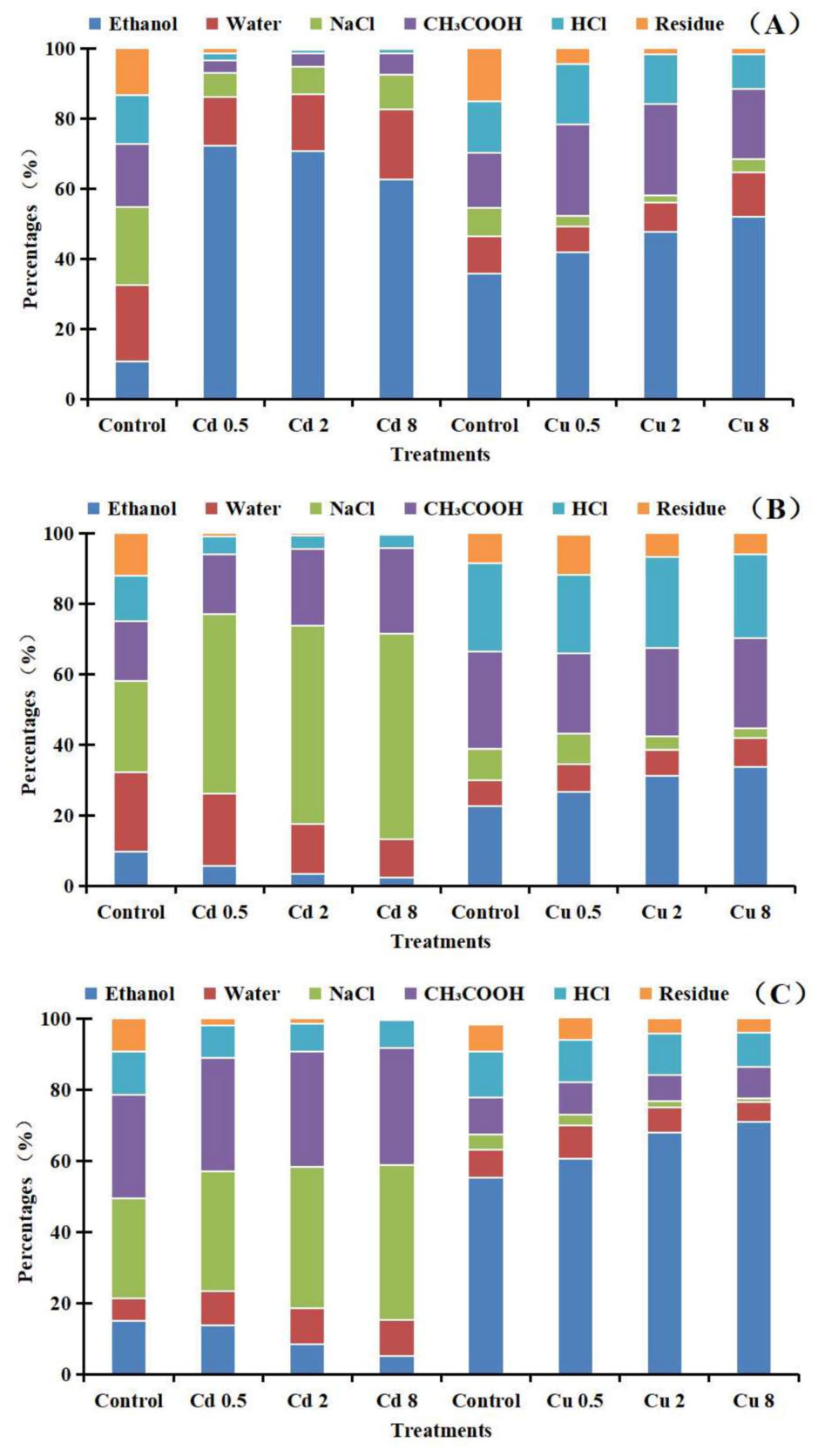
Cd increased. The transformation of chemical form was the main detoxification mechanism of B. pilosa seedling stems coping with Cd stress. But ethanol-extracted Cu remained unchanged in the stems. As shown from Fig. 4C, the proportion of Cd chemical forms in leaves was ordered as follows: NaCl (39%) > CH3COOH (32%) > Water (9.9%) > Ethanol (9.1%) > HCl (8.2%) > Residue (1.4%), and the proportion of Cd extracted with NaCl or CH3COOH elevated with Cd stress increased from 0.5 to 8 mg L−1. By contrast, ethanol extract the highest Cu concentration in all the Cu extracts, and the proportion of Cu extracted with other agents was relatively low. The results indicated that the detoxification mechanism of B. pilosa leaves was by means of elevating the proportion of NaCl-extracted Cd and lowering the ethanol-extracted Cd. But the leaves had no detoxification ability to Cu.
Cd比例升高。化学形态的转化是B. pilosa幼苗茎应对Cd胁迫的主要解毒机制,而茎中乙醇提取态Cu保持不变。如图4C所示,叶中各化学形态Cd的比例依次为:NaCl提取态(39%)> CH3COOH提取态(32%)> 水提取态(9.9%)> 乙醇提取态(9.1%)> HCl提取态(8.2%)> 残渣态(1.4%),且当Cd胁迫由0.5增至8 mg L−1时,NaCl或CH3COOH提取态Cd比例升高。相比之下,在所有Cu提取物中乙醇提取的Cu浓度最高,其他提取剂提取的Cu比例相对较低。结果表明,B. pilosa叶片的解毒机制是提高NaCl提取态Cd比例并降低乙醇提取态Cd比例,但叶片对Cu不具备解毒能力。
5Discussion讨论
Dai et al. (2017) found that the Cd concentration in B. pilosa shoots was 405.91 mg kg−1 after four weeks of growth in soils with Cd concentration at 64 mg/ kg, and the translocation and enrichment factors were 1.14 and 6.33, respectively. Cd concentration in shoots was 1652 mg kg−1, when the plants were cultivated in nutrient solutions with a Cd concentration of 64 mg/kg, and the translocation and enrichment factors were 1.49 and 25.81, respectively. The shoot Cd concentrations in B. pilosa in this study were less than those reported by Dai et al. (2017). This may be due to phosphate co-precipitation forming in the root apoplast. Liu et al. (2019) identified Lantana camara L. as a Cd hyperaccumulator using dose gradient experiments and field tests. These authors reported that the seedlings were grown in the contaminated soils with Cd level lower than 100 mg kg−1, and the plants showed high Cd tolerance, without obvious damage and reduction in shoot biomass compared to controls. Importantly, the bioaccumulation and translocation factors were all > 1, and the Cd concentration in the shoots was also higher than 100 mg kg−1 in all treatments. Xv et al. (2020) found that Cu enrichment in Sedum alfredii mainly occurred in the roots, with only a minimal amount of Cu translocation to the stem. The high tolerance of S. alfredii to Cu resulted from homeostasis in root cells, and vascular tissues were the dominant location of Cu restriction. Our research suggested that B. pilosa exhibited some basic characteristics of Cd hyperaccumulator in hydroponic culture and shoot Cd concentration was up to 100 mg kg−1 in the 0.5 mg L−1 Cd treatment. The TF of B. pilosa was 1.26, and the roots showed high tolerance to Cd, the results here might be different to other researchers, which were originated from a different ecotype of B. pilosa or under different growth conditions. However, this species did not show Cu hyperaccumulator or accumulator properties, because the TFs were all < 1 in Cu treatments. In addition, most Cu concentrated in the roots, and root biomass decreased under Cu stress conditions.
Dai et al. (2017)发现,B. pilosa在Cd浓度为64 mg/ kg的土壤中生长4周后,地上部Cd浓度为405.91 mg kg−1,转运系数和富集系数分别为1.14和6.33;在Cd浓度为64 mg/kg的营养液中培养时,地上部Cd浓度为1652 mg kg−1,转运系数和富集系数分别为1.49和25.81。本研究中B. pilosa地上部Cd浓度低于Dai et al. (2017)的报道值,这可能是由于根质外体中形成了磷酸盐共沉淀。Liu et al. (2019)通过剂量梯度试验和田间试验确定Lantana camara L.为Cd超富集植物。上述作者报道,幼苗生长在Cd含量低于100 mg kg−1的污染土壤中时,植株表现出较高的Cd耐受性,与对照相比无明显损伤,地上部生物量也未减少。重要的是,其生物富集系数和转运系数均 > 1,且所有处理中地上部Cd浓度均高于100 mg kg−1。Xv et al. (2020)发现,Sedum alfredii对Cu的富集主要发生在根中,仅有极少量Cu转运至茎。S. alfredii对Cu的高耐受性源于根细胞内的稳态,维管组织是Cu受限的主要部位。本研究表明,B. pilosa在水培条件下表现出Cd超富集植物的一些基本特征,在0.5 mg L−1 Cd处理中地上部Cd浓度可达100 mg kg−1。B. pilosa的TF为1.26,且根系对Cd表现出较高耐受性;本研究结果可能与其他研究者不同,这可能源于B. pilosa生态型不同或生长条件不同。然而,该物种未表现出Cu超富集植物或富集植物的特性,因为Cu处理中的TF均 < 1。此外,大部分Cu集中于根中,且在Cu胁迫条件下根生物量降低。
Plants produce metal-binding proteins such metallothioneins and phytochelatins that chelate and sequester heavy metals (REF). These proteins bind to metals and prevent them from interfering with important physiological processes thereby reducing their toxicity (Fahad et al. 2024, 2021). Mwamba et al. (2016) found that the main chemical forms of Cd integration in rapeseed (Brassica napus) roots were pectates and proteins and phosphate-bound Cu was the dominant fraction in the cell walls. Bora and Sarma (2021) analyzed the FTIR spectrum of Ceratopteris pteridoides root samples and observed strong and broad peaks in the mid-infrared region of 3500 ~ 3200 cm−1, 1700 ~ 1600 cm−1, 1500 ~ 1400 cm−1, 1300 ~ 1200 cm−1 and 800 ~ 400 cm−1. These peaks represented N–H or O–H, N–H, C-H, P = O or C-O or C-O-P, C-O–H or C-S functional groups, which played significant roles in Cd binding and corresponded to lignin, cellulose, proteins, lipids, phospholipids or nucleic acids, disulfide bonds and other metabolites. The latter authors also found that the mechanism of Cd tolerance in Ceratopteris pteridoides roots was based on the accumulation of electron-dense material on the cell walls. Wu et al. (2020) used FTIR analysis to investigate the mechanisms of Cd resistance in rapeseed (Brassica napus) shoots with the addition of boron (B) and suggested that the increase in pectin, protein, carbohydrate and cellulose contents in the cell walls alleviated Cd toxicity. Su et al. (2017) found that amide, hydroxyl, carbonyl and thiol functional groups were involved in Cd accumulation and uptake in duckweed (Spirodela polyrhiza) roots on the basis of FTIR spectrometry analysis, and reported that Cd stress could induce the accumulation of soluble sugars. Dumont et al. (2022) studied the toxic effects of nano-CuO on Myriophyllum spicatum seedlings and suggested that the composition and structure of biomacromolecules (such as proteins, carbohydrates, phenols and cellulosic compounds) in the plant samples markedly changed under different nano-CuO treatments (from 5 to 70 mg L−1). Rana et al. (2018) argued that the characteristic peaks at 1385 ~ 1394 cm−1represented stretching vibrations of oxygen and hydrogen, indicating changes in cellulose contents in orchardgrass (Dactylis glomerata L.) leaf tissue; on the other hand, the absorbance peaks at 1373 ~ 1375 cm−1 were shown to represent suberinlike aliphatic compounds in kentucky bluegrass (Poa pratensis L.) leaves. Sawalha et al. (2007) used FTIR to investigate the identity of functional groups involved in Cd binding in different tissues of saltbush (Atriplex canescens) shoots after acidic methanol modification and found that the carboxyl group was the dominant one in Cd binding sites. Zhang et al. (2021) found that the detoxification mechanisms of annual fleabane (Erigeron annuus) in response to 200 μmol L−1 Cd stress involved the secretion of dissolved organic matter (DOM) by roots, and DOM mainly consisted of several functional groups, such as aromatic structures, tannins and carbohydrates, which played important roles in stress management. Yu et al. (2020) analyzed the FTIR spectral information of root, stem and leaf samples from Conyza canadensis (L.) Cronq., and found that the shapes of characteristic peaks remained unchanged in Cd treatments and controls, but the absorbance intensity of the roots was stronger under high Cd concentration conditions, indicating that large quantities of proteins and amino acids were produced as nitrogen sources to manage Cd toxicity and sustain intracellular environments. Lan et al. (2019) investigated Cd distribution and accumulation in the roots, stems and leaves of the hyperaccumulator Microsorum pteropus and found that Cd was mainly sequestrated and stored in the roots and leaf cell wall fraction; in addition to the cell wall, Cd was also found in the stem cytoplasm fraction. He et al. (2020) analyzed subcellular fractions of different tissues from castor (Ricinus communis) seedlings using FTIR and discovered that functional groups (carboxyl, hydroxyl, amide and amino groups) provided a large number of sites for Cd binding on the cell wall. Our research also suggested that typical functional groups (e.g., -OH; -CH2 and -CH(CH3)2)
植物可产生金属硫蛋白和植物螯合肽等金属结合蛋白,以螯合并隔离重金属(REF)。这些蛋白质与金属结合,阻止其干扰重要的生理过程,从而降低其毒性(Fahad et al. 2024, 2021)。Mwamba et al. (2016)发现,油菜(Brassica napus)根中Cd结合的主要化学形态为果胶酸盐和蛋白质,而磷酸盐结合态Cu是细胞壁中的主要组分。Bora and Sarma (2021)分析了Ceratopteris pteridoides根样品的FTIR光谱,在中红外区域的3500 ~ 3200 cm−1、1700 ~ 1600 cm−1、1500 ~ 1400 cm−1、1300 ~ 1200 cm−1和800 ~ 400 cm−1处观察到强而宽的吸收峰。这些峰分别代表N–H或O–H、N–H、C-H、P = O或C-O或C-O-P、C-O–H或C-S官能团,它们在Cd结合中发挥重要作用,对应于木质素、纤维素、蛋白质、脂类、磷脂或核酸、二硫键及其他代谢物。上述作者还发现,Ceratopteris pteridoides根的Cd耐受机制基于细胞壁上电子致密物质的积累。Wu et al. (2020)利用FTIR分析研究了添加硼(B)条件下油菜(Brassica napus)地上部的抗Cd机制,认为细胞壁中果胶、蛋白质、碳水化合物和纤维素含量的增加缓解了Cd毒性。Su et al. (2017)基于FTIR光谱分析发现,酰胺、羟基、羰基和巯基官能团参与了紫萍(Spirodela polyrhiza)根对Cd的积累和吸收,并报道Cd胁迫可诱导可溶性糖的积累。Dumont et al. (2022)研究了纳米CuO对Myriophyllum spicatum幼苗的毒性效应,认为在不同纳米CuO处理(5至70 mg L−1)下,植物样品中生物大分子(如蛋白质、碳水化合物、酚类和纤维素类化合物)的组成和结构发生了明显变化。Rana et al. (2018)认为,1385 ~ 1394 cm−1处的特征峰代表氧和氢的伸缩振动,指示鸭茅(Dactylis glomerata L.)叶组织中纤维素含量的变化;另一方面,1373 ~ 1375 cm−1处的吸收峰被证明代表草地早熟禾(Poa pratensis L.)叶片中的类木栓质脂肪族化合物。Sawalha et al. (2007)利用FTIR研究了经酸性甲醇修饰后四翅滨藜(Atriplex canescens)地上部不同组织中参与Cd结合的官能团,发现羧基是Cd结合位点中的主要官能团。Zhang et al. (2021)发现,一年蓬(Erigeron annuus)响应200 μmol L−1 Cd胁迫的解毒机制涉及根系分泌溶解性有机质(DOM),DOM主要由芳香结构、单宁和碳水化合物等若干官能团组成,在胁迫应对中发挥重要作用。Yu et al. (2020)分析了Conyza canadensis (L.) Cronq.根、茎和叶样品的FTIR光谱信息,发现Cd处理与对照中特征峰的形状保持不变,但在高Cd浓度条件下根的吸收强度更强,表明植物产生了大量蛋白质和氨基酸作为氮源,以应对Cd毒性并维持细胞内环境。Lan et al. (2019)研究了超富集植物Microsorum pteropus根、茎和叶中Cd的分布与积累,发现Cd主要被隔离并储存于根和叶的细胞壁组分中;除细胞壁外,茎的细胞质组分中也检测到Cd。He et al. (2020)利用FTIR分析了蓖麻(Ricinus communis)幼苗不同组织的亚细胞组分,发现官能团(羧基、羟基、酰胺基和氨基)为细胞壁上的Cd结合提供了大量位点。本研究也表明,典型官能团(如-OH、-CH2和-CH(CH3)2)
and corresponding biochemical regions, such as acid amides and lipids were involved in the co-precipitation and complexation of B. pilosa root cells as a response to Cd stress.
及相应的生化区域(如酸酰胺和脂类)参与了B. pilosa根细胞响应Cd胁迫的共沉淀和络合作用。
Pan et al. (2019) discovered that manganese (Mn) was isolated in cell wall and vacuole of pink plumepoppy (Macleaya cordata). In addition, most Mn was sequestered in low-toxicity compound such as oxalates, pectates, phosphates and protein. Teng et al. (2021) suggested that the vacuoles of S. nigrum leaf cells were main locations for Cd storage, and the proportion of vacuolar Cd in protoplasts was up to 83.37% under 50 μM Cd treatments. In addition, the chelation state Cd was the dominated chemical form in protoplast. Luo et al. (2024) demonstrated that More than 50% of Cd was trapped in cell walls from leaves of reed fescue (Festuca arundinacea). The cell wall binding Cd was a key pathway for detoxification. The cell wall served as a protective barrier against Cd penetration into plant cells. Some plants changed the makeup of their cell walls, increased the binding of Cd to pectins, hemicelluloses, and other structural elements (Ahmad et al. 2022; Irfan et al. 2021; Fahad et al. 2020). Cd-induced changed in cell wall composition, increased lignification and changed in polysaccharide content, contributed to a physical barrier that restricted the entry of Cd ions into plant cells (Song et al. 2024). An et al. (2023) verified that Cd was mainly accumulated in Platycodon grandiflorum roots, and was predominantly sequestered in cell wall. The chemical state of Cd was characterized by low mobility and toxicity, and the proportion of Cd in cytoplasm increased under Cd stress. The accumulation and detoxification mechanism of Cd by P. grandiflorum were root retention, cell wall deposition, vacuole isolation, low mobility and toxicity transformation. Wu et al. (2016) showed that Cd proportion in cell wall of S. matsudana roots was 53% under 10 μM Cd stress, and concluded that cell wall of roots might serve as the first barrier to bind and reduce free Cd ions entering the cytoplasm. The proportion of ethanol-extracted and water-extracted Cd were relatively low, and the proportion of NaCl-extracted Cd was 42% with 10 μM Cd treatment. Xin et al. (2014) compared the difference of Cd uptake and translocation between low and high accumulation cultivars of hot pepper by measuring the subcellular distributions and chemical forms of Cd, and concluded that the cytoplasm stored the majority of Cd in the roots of both varieties, and a minimal amount of Cd existed in organelle fraction. These researches were basically consistent with our results. Our results showed that most of Cd was mainly concentrated in cytoplasm of B. pilosa roots, and sequestered in cell wall of stems and leaves to cope with stress. The detoxification mechanism of B. pilosa was to elevate the proportion of NaCl-extracted Cd and lower ethanol-extracted Cd.
Pan et al. (2019)发现,锰(Mn)被隔离于博落回(Macleaya cordata)的细胞壁和液泡中;此外,大部分Mn以草酸盐、果胶酸盐、磷酸盐和蛋白质等低毒性化合物形式被封存。Teng et al. (2021)指出,S. nigrum叶片细胞的液泡是Cd储存的主要部位,在50 μM Cd处理下,原生质体中液泡Cd所占比例高达83.37%;此外,螯合态Cd是原生质体中的主要化学形态。Luo et al. (2024)证实,苇状羊茅(Festuca arundinacea)。叶片中超过50%的Cd被截留于细胞壁中,细胞壁结合Cd是其解毒的关键途径。细胞壁可作为阻止Cd进入植物细胞的保护屏障。一些植物通过改变细胞壁组成,增强Cd与果胶、半纤维素及其他结构组分的结合(Ahmad et al. 2022; Irfan et al. 2021; Fahad et al. 2020)。Cd诱导的细胞壁组成变化、木质化增强以及多糖含量改变,共同形成了限制Cd离子进入植物细胞的物理屏障(Song et al. 2024)。An et al. (2023)证实,Cd主要积累于桔梗(Platycodon grandiflorum)根部,且主要被封存于细胞壁中;Cd的化学形态表现为低迁移性和低毒性,而在Cd胁迫下细胞质中Cd的比例有所增加。P. grandiflorum对Cd的积累与解毒机制包括根部滞留、细胞壁沉积、液泡隔离以及向低迁移性、低毒性形态的转化。Wu et al. (2016)研究表明,在10 μM Cd胁迫下,S. matsudana根系细胞壁中Cd所占比例为53%,并认为根系细胞壁可能是结合并减少进入细胞质的游离Cd离子的第一道屏障;在10 μM Cd处理下,乙醇提取态和水提取态Cd的比例相对较低,而NaCl提取态Cd的比例为42%。Xin et al. (2014)通过测定Cd的亚细胞分布和化学形态,比较了辣椒低积累与高积累品种在Cd吸收和转运上的差异,认为两个品种根系中的Cd大部分储存于细胞质中,而细胞器组分中的Cd含量极少。上述研究与本研究结果基本一致。本研究结果表明,大部分Cd主要富集于B. pilosa根系的细胞质中,而在茎和叶中则被封存于细胞壁中,以应对胁迫。B. pilosa的解毒机制是提高NaCl提取态Cd的比例并降低乙醇提取态Cd的比例。
6Conclusions结论
Our work showed that the key compounds associated with Cd hyperaccumulation in B. pilosa were alcohols, phenols, acid amides and lipids by using FTIR, focusing specifically on differences in functional group characteristics. The study further confirmed that B. pilosa was a Cd hyperaccumulator, but not a Cu hyperaccumulator. The FTIR results indicated that the characteristic peaks corresponded to the following functional groups and biochemical regions: phenolic and alcohol -OH groups, -CH2 groups, acid amides, -CH(CH3)2 groups and fatty acids. The amount of these functional groups increased with increasing Cd stress concentrations, and corresponding macromolecules in cells in the stems/roots of B. pilosa participated in response to Cd stress stimuli. Especially, the absorbance peak intensity at 1385 cm−1 in root spectra increased gradually with increasing Cd stress, but decreased under Cu stress. The results indicated that a number of -CH(CH3)2 groups were produced to combine with Cd in root cell surface. In contrast, the amount of -CH(CH3)2 groups was reduced with increasing Cu stress, the amount of -CH(CH3)2 groups and protein with acid amides were raising in cells in the stems of B. pilosa under Cd treatments. Results also revealed that most of Cd was mainly sequestered in cytoplasm and cell walls of B. pilosa roots to cope with Cd stress. Cu was only trapped in cell walls of root tissues, and majority of Cd/Cu was sequestered in cell walls of B. pilosa stems and leaves to cope with stress. The detoxification of B. pilosa was completed by means of elevating the proportion of NaCl-extracted Cd and lowering ethanol-extracted Cd. But there were no significant changes for the proportion of ethanol-extracted Cu. The hyperaccumulating and detoxification mechanisms of B.
本研究利用FTIR技术,重点关注官能团特征的差异,结果表明,与B. pilosaCd超富集相关的关键化合物为醇类、酚类、酰胺类和脂类。本研究进一步证实,B. pilosa是Cd超富集植物,而非Cu超富集植物。FTIR结果表明,特征峰对应以下官能团及生化区域:酚类和醇类的-OH基团、-CH2基团、酰胺、-CH(CH3)2基团以及脂肪酸。这些官能团的数量随Cd胁迫浓度的升高而增加,B. pilosa茎/根细胞中的相应大分子参与了对Cd胁迫刺激的响应。尤其是,根系光谱中1385 cm−1处的吸收峰强度随Cd胁迫的增强而逐渐升高,但在Cu胁迫下则降低。结果表明,根细胞表面产生了大量-CH(CH3)2基团以结合Cd。相反,-CH(CH3)2基团的数量随Cu胁迫的增强而减少;在Cd处理下,B. pilosa茎细胞中-CH(CH3)2基团及含酰胺的蛋白质数量增加。结果还表明,大部分Cd主要被封存于B. pilosa根系的细胞质和细胞壁中,以应对Cd胁迫。Cu仅被截留于根组织的细胞壁中,而在B. pilosa茎和叶中,大部分Cd/Cu被封存于细胞壁中以应对胁迫。B. pilosa通过提高NaCl提取态Cd的比例并降低乙醇提取态Cd的比例完成解毒,但乙醇提取态Cu的比例无显著变化。B.
pilosa might be regulation of the corresponding macromolecules (such as alcohols and phenols, proteins and lipids, etc.) in cells, Cd immobilization and compartmentalization in cytoplasm and cell walls, and transformation of Cd chemical forms. The response to Cd stress of B. pilosa represented a complex physiological and biochemical metabolic process. So the research of Cd subcellular distributions and chemical forms should be combined with various omics level methods such as genomics, transcriptomics, proteomics and metabolomics to study the relationships between genes, proteins and metabolites to improve the understanding of B. pilosa responses to Cd stress.
pilosa的超富集与解毒机制可能包括:调控细胞中相应的大分子(如醇类和酚类、蛋白质和脂类等),将Cd固定并区隔化于细胞质和细胞壁中,以及Cd化学形态的转化。B. pilosa对Cd胁迫的响应是一个复杂的生理生化代谢过程。因此,Cd亚细胞分布与化学形态的研究应与基因组学、转录组学、蛋白质组学和代谢组学等多种组学方法相结合,研究基因、蛋白质与代谢产物之间的关系,以加深对B. pilosa响应Cd胁迫的认识。
Author contribution Siqi Wang: Data processing, Writing – original draft & detection. Jiayi Bai: Resources, cultivation & detection. Huiping Dai: Data curation, Formal analysis, Writing – review & editing. Jie Zhan: Validation, Writing – review & editing. Liping Ren: Detection & data processing. Brett H. Robinson: Validation, Writing – review & editing. Chengzhi Jiang: Detection & data processing. Shuang Cui: Detection & data processing. Lidia Skuza: Validation, Writing – review & editing. Shuhe Wei: Conceptualization, Methodology, Project administration, Supervision.
作者贡献 Siqi Wang:数据处理,撰写—初稿及检测。Jiayi Bai:资源,培养及检测。Huiping Dai:数据整理,形式分析,撰写—审阅与编辑。Jie Zhan:验证,撰写—审阅与编辑。Liping Ren:检测及数据处理。Brett H. Robinson:验证,撰写—审阅与编辑。Chengzhi Jiang:检测及数据处理。Shuang Cui:检测及数据处理。Lidia Skuza:验证,撰写—审阅与编辑。Shuhe Wei:构思,方法,项目管理,指导。
Funding This work was supported by Sanqin Talents, Shaanxi Provincial First-class Team– " Contaminated Soil Remediation and Resource Utilization Innovation Team at Shaanxi University of Technology", the National Natural Science Foundation of China (32471703), Shaanxi University of Technology Research Ability Enhancement Project (SLGNL202405), Qin Chuangyuan "Scientists + Engineers" Team Construction in Shaanxi Province (2024QCY- KXJ-104), the project of Foreign Experts Bureau of China (S20240182, 2025WZ-YBXM-19, 2025WZ-YBXM-29), Shaanxi Province City-University Co-Construction Project (SXZJ-2301), Qinba Bioremediation and Resource Development Research Innovation and Introduction Base (2025YZ- YIPT-41; S2022-ZC-GXYZ-0029), the program financed by Poland Minister of Science under the "Regional Excellence Initiative" for 2024–2027, the General Program of the National Natural Science Foundation of China (41671324), the General Program from the Education Department of Liaoning Province (LJKMZ20220595). Basic scientific research project of Liaoning Provincial Department of Education (General Project) (JYTMS20230179).
基金资助 本研究得到以下项目资助:三秦人才、陕西省一流团队——“陕西理工大学污染土壤修复与资源化利用创新团队”,国家自然科学基金(32471703),陕西理工大学科研能力提升项目(SLGNL202405),陕西省秦创原“科学家+工程师”队伍建设(2024QCY- KXJ-104),国家外国专家局项目(S20240182, 2025WZ-YBXM-19, 2025WZ-YBXM-29),陕西省市校共建项目(SXZJ-2301),秦巴生物修复与资源开发研究创新引智基地(2025YZ- YIPT-41; S2022-ZC-GXYZ-0029),波兰科学部长“区域卓越计划”(2024–2027)资助项目,国家自然科学基金面上项目(41671324),辽宁省教育厅面上项目(LJKMZ20220595),辽宁省教育厅基本科研项目(面上项目)(JYTMS20230179)。
Data availability Data will be made available on request.
数据可用性 数据可应要求提供。
Declarations声明
Competing interest The authors declare that they have no known competing financial interests or personal relationships that could have appeared to influence the work reported in this paper.
利益冲突 作者声明,不存在任何可能影响本文所报道工作的已知经济利益冲突或个人关系。
7References参考文献
Ahmad W, Khan A, Zeeshan M, Ahmad I, Adnan M, Fahad S, Solaiman Z (2022) Relative efficiency of biochar particles of different sizes for immobilising heavy metals and improving soil properties. Crop Pasture Sci 74:112–120
An J, Wang X, Jing Y, Huang J, Wang Q, Zhang G, Gao J, Peng L, Huang W, Yan Y (2023) Subcellular distribution and chemical forms of cadmium in the medicine food homology plant Platycodon grandiflorum (Jacq.) A.DC. Phyton (0031-9457) 92(5):1405–1420
Bora MS, Sarma KP (2021) Anatomical and ultrastructural alterations in Ceratopteris pteridoides under cadmium stress: A mechanism of cadmium tolerance. Ecotoxicol Environ Saf 218:112285
Cai FJ, Wang L, Zhao W, Tian JL, Kong DG, Liu Q, Sun XH, Zhou HL (2022) Phytochemical and chemotaxonomic investigations on the whole herbs of Bidens procera L.C.Xu ex X.W.Zheng. Biochem Syst Ecol 101:104395 Chow YY, Ting ASY (2019) Influence of fungal infection on plant tissues: FTIR detects compositional changes to plant cell walls. Fungal Ecol 37:38–47
Dai H, Wei S, Twardowska I, Han R, Xu L (2017) Hyperaccumulating potential of Bidens pilosa L. for Cd and elucidation of its translocation behavior based on cell membrane permeability. Environ Sci Pollut Res 24(29):23161–23167
Dumont ER, Elger A, Azéma C, Michel HC, Surble S, Larue C (2022) Cutting-edge spectroscopy techniques highlight toxicity mechanisms of copper oxide nanoparticles in the aquatic plant Myriophyllum spicatum. Sci Total Environ 803:150001
Fahad S, Hasanuzzaman M, Alam M, Ullah H, Saeed M, Khan IA, Adnan M (eds) (2020) Environment, climate, plant and vegetation growth. Springer International Publishing, Berlin/Heidelberg, Germany, pp 397–419
Fahad S, Saud S, Yajun C, Chao W, Depeng W (Eds.) (2021) Abiotic stress in plants. Intech Open United Kingdom 2021.
Fahad S, Saud S, Nawaz T, Gu L, Ahmad M, Zhou R (Eds.) (2024) Environment, Climate, Plant and Vegetation Growth (2nd edition). Springer Nature Switzerland AG 2024.
Feng Y, Wang Q, Meng Q, Liu Y, Pan F, Luo S, Wu Y, Ma L, Yang X (2019) Chromosome doubling of Sedum alfredii Hance: A novel approach for improving phytoremediation efficiency. J Environ Sci 86:87–96
Hamid Y, Tang L, Yaseen M, Hussain B, Zehra A, Aziz MZ, He Z, Yang X (2019) Comparative efficacy of organic and inorganic amendments for cadmium and lead immobilization in contaminated soil under rice-wheat cropping system. Chemosphere 214:259–268
He C, Zhao Y, Wang F, Oh K, Zhao Z, Wu C, Zhang X, Chen X, Liu X (2020) Phytoremediation of soil heavy metals (Cd and Zn) by castor seedlings: Tolerance, accumulation and subcellular distribution. Chemosphere 252:126471
Heredia B, Tapia R, Young BJ, Hasuoka P, Pacheco P, Roqueiro G (2022) Phytoextraction of Cu, Cd, Zn and As in four shrubs and trees growing on soil contaminated with mining waste. Chemosphere 308:136146
Irfan M, Mudassir M, Khan MJ, Dawar KM, Muhammad D, Mian IA, Ali W, Fahad S, Saud S, Hayat Z, Nawaz T, Khan SA, Alam S, Ali B, Banout J, Ahmed S, Mubeen S, Danish S, Datta R, Elgorban AM, Dewil R (2021) Heavy metals immobilization and improvement in maize (Zea mays L.) growth amended with biochar and compost. Sci Rep 11(1):18416
Lan XY, Yan YY, Yang B, Li XY, Xu FL (2019) Subcellular distribution of cadmium in a novel potential aquatic hyperaccumulator-Microsorum pteropus. Environ Pollut 248:1020–1027
Li Z, Yang Y, Guan W, Yu H, Zou L, Cui J, Teng Y (2023) Insight into the subcellular mechanism of maximizing Cd accumulation in hyperaccumulator Solanum nigrum L. under the action of biodegradable chelating agent. Environ Exp Bot 207:105226
Liu S, Ali S, Yang R, Tao J, Ren B (2019) A newly discovered Cd-hyperaccumulator Lantana camara L. J Hazard Mater 371:233–242
Liu X, Renard CM, Bureau S, Le Bourvellec C (2021) Revisiting the contribution of ATR-FTIR spectroscopy to characterize plant cell wall polysaccharides. Carbohyd Polym 262:117935 Luo J, Feng S, Li M, He Y, Deng Y, Cao M (2024) Effect of magnetized water irrigation on Cd subcellular allocation and chemical forms in leaves of Festuca arundinacea during phytoremediation. Ecotoxicol Environ Saf 277(116376):455–462
Mwamba TM, Li L, Gill RA, Islam F, Nawaz A, Ali B, Farooq MA, Lwalaba JL, Zhou WJ (2016) Differential subcellular distribution and chemical forms of cadmium and copper in Brassica napus. Ecotoxicol Environ Saf 134:239–249
Omara T, Kagoya S, Openy A, Omute T, Ssebulime S, Kiplagat KM, Bongomin O (2020) Antivenin plants used for treatment of snakebites in Uganda: ethnobotanical reports and pharmacological evidences. Trop Med Health 48(1):1–16
Pan F, Meng Q, Wang Q, Luo S, Chen B, Khan KY, Yang X, Feng Y (2016) Endophytic bacterium Sphingomonas SaMR12 promotes cadmium accumulation by increasing glutathione biosynthesis in Sedum alfredii Hance. Chemosphere 154:358–366
Pan G, Zhang H, Liu W, Liu P (2019) Integrative study of subcellular distribution, chemical forms, and physiological responses for understanding manganese tolerance in the herb Macleaya cordata (papaveraceae). Ecotoxicol Environ Saf 181:455–462
Pasricha S, Mathur V, Garg A, Lenka S, Verma K, Agarwal S (2021) Molecular mechanisms underlying heavy metal uptake, translocation and tolerance in hyperaccumulatorsan analysis: Heavy metal tolerance in hyperaccumulators. Environ Challenges 4:100197
Rana R, Herz K, Bruelheide H, Dietz S, Haider S, Jandt U, Pena R (2018) Leaf Attenuated Total Reflection Fourier Transform Infrared (ATR-FTIR) biochemical profile of grassland plant species related to land-use intensity. Ecol Indic 84:803–810
Sawalha MF, Peralta-Videa JR, Saupe GB, Dokken KM, Gardea-Torresdey JL (2007) Using FTIR to corroborate the identity of functional groups involved in the binding of Cd and Cr to saltbush (Atriplex canescens) biomass. Chemosphere 66(8):1424–1430
Sharma S, Uttam KN (2017) Rapid analyses of stress of copper oxide nanoparticles on wheat plants at an early stage by laser induced fluorescence and attenuated total reflectance Fourier transform infrared spectroscopy. Vib Spectrosc 92:135–150
Sharma S, Uttam KN (2019) Non-destructive and rapid interrogation of biochemical response of the leaves of wheat seedlings towards Al2O3 nanoparticles stress using attenuated total reflectance Fourier transform infrared spectroscopy. Vib Spectrosc 100:142–151
Song J, Sun Z, Saud S, Fahad S, Nawaz T (2024) Exploring the deleterious effects of heavy metal cadmium on antioxidant defense and photosynthetic pathways in higher plants. Plant Stress 100716
Su C, Jiang Y, Li F, Yang Y, Lu Q, Zhang T, Hu D, Xu Q (2017) Investigation of subcellular distribution, physiological, and biochemical changes in Spirodela polyrhiza as a function of cadmium exposure. Environ Exp Bot 142:24–33
Sun Y, Zhou Q, Wang L, Liu W (2009) Cadmium tolerance and accumulation characteristics of Bidens pilosa L. as a potential Cd-hyperaccumulator. J Hazard Mater 161(2–3):808–814 Suresh S, Karthikeyan S, Jayamoorthy K (2016) FTIR and multivariate analysis to study the effect of bulk and nano copper oxide on peanut plant leaves. J Sci-Adv Mater Dev 1(3):343–350
Teng Y, Yu A, Tang YM, Jiang ZY, Guan WJ, Li ZS, Yu HY, Zou LY (2021) Visualization and quantification of cadmium accumulation, chelation and antioxidation during the process of vacuolar compartmentalization in the hyperaccumulator plant Solanum nigrum L. Plant Sci 310:110961
Tolmacheva AA, Rogozhin EA, Deryabin DG (2014) Antibacterial and quorum sensing regulatory activities of some traditional Eastern-European medicinal plants. Acta Pharmaceut 64(2):173–186
Wang S, Dai H, Skuza L, Chen Y, Wei S (2022) Difference in Cd2+ flux around the root tips of different soybean (Glycine max L.) cultivars and physiological response under mild cadmium stress. Chemosphere 297:134120
Wei S, Zhou Q (2008) Screen of Chinese weed species for cadmium tolerance and accumulation characteristics. Int J Phytoremediat 10(6):584–597
Wei S, Zhou Q, Zhan J, Wu Z, Sun T, Lyubu Y, Prasad MNV (2010) Poultry manured Bidens tripartite L. extracting Cd from soil-potential for phytoremediating Cd contaminated soil. Bioresource Technol 101(22):8907–8910
Wei H, Huang M, Quan G, Zhang J, Liu Z, Ma R (2018) Turn bane into a boon: Application of invasive plant species to remedy soil cadmium contamination. Chemosphere 210:1013–1020
Wu H, Wang J, Li B, Ou Y, Wang J, Shi Q, Jiang W, Liu D, Zou J (2016) Salix matsudana koidz tolerance mechanisms to cadmium: Uptake and accumulation, subcellular distribution, and chemical forms. Pol J Environ Stud 25(4):1739–1747
Wu X, Song H, Guan C, Zhang Z (2020) Boron mitigates cadmium toxicity to rapeseed (Brassica napus) shoots by relieving oxidative stress and enhancing cadmium chelation onto cell walls. Environ Pollut 263:114546
Xin J, Huang B, Dai H, Liu A, Zhou W, Liao K (2014) Characterization of cadmium uptake, translocation, and distribution
in young seedlings of two hot pepper cultivars that differ in fruit cadmium concentration. Environ Sci Pollut Res 21:7449–7456
Xu M, Lin Y, da Silva EB, Cui Q, Gao P, Wu J, Ma LQ (2022) Effects of copper and arsenic on their uptake and distribution in As-hyperaccumulator Pteris vittata. Environ Pollut 300:118982
Xv L, Ge J, Tian S, Wang H, Yu H, Zhao J, Lu L (2020) A Cd/Zn Co-hyperaccumulator and Pb accumulator, Sedum alfredii, is of high Cu tolerance. Environ Pollut 263:114401
Yang Q, Shohag MJI, Feng Y, He Z, Yang X (2017) Transcriptome comparison reveals the adaptive evolution of two contrasting ecotypes of Zn/Cd hyperaccumulator Sedum alfredii hance. Front Plant Sci 8:425
Yu S, Sheng L, Zhang C, Deng H (2018) Physiological response of Arundo donax to cadmium stress by Fourier transform infrared spectroscopy. Spectrochim Acta A 198:88–91
Yu S, Sheng L, Mao H, Huang X, Luo L, Li Y (2020) Physiological response of Conyza canadensis to cadmium stress monitored by Fourier transform infrared spectroscopy and cadmium accumulation. Spectrochim Acta A 229:118007
Yu H, Yang A, Wang K, Li Q, Ye D, Huang H, Zhang X, Wang Y, Zheng Z, Li T (2021) The role of polysaccharides functional groups in cadmium binding in root cell wall of a cadmiumsafe rice line. Ecotoxicol Environ Saf 226:112818
Yu F, Tang S, Shi X, Liang X, Liu K, Huang Y, Li Y (2022) Phytoextraction of metal (loid) s from contaminated soils by six plant species: A field study. Sci Total Environ 804:150282
Yu SH, Deng HP, Zhang B, Liu ZX, Lin JJ, Sheng L, Qi JS (2022) Physiological response of Vetiveria Zizanioides to cadmium stress revealed by Fourier transform infrared spectroscopy. Spectrosc Lett 55(3):157–165
Zeng HY, Chen LH, Yang Y, Deng X, Zhou XH, Zeng QR (2019) Basal and foliar treatment using an organic fertilizer amendment lowers cadmium availability in soil and cadmium uptake by rice on field micro-plot experiment planted in contaminated acidic paddy soil. Soil Sediment Contam 28(1):1–14
Zhang H, Heal K, Zhu X, Tigabu M, Xue Y, Zhou C (2021) Tolerance and detoxification mechanisms to cadmium stress by hyperaccumulator Erigeron annuus include molecule synthesis in root exudate. Ecotoxicol Environ Saf 219:112359
Zou J, Wang Y, Wang S, Shang X (2023) Ca alleviated Cd-induced toxicity in Salix matsudana by affecting Cd absorption, translocation, subcellular distribution, and chemical forms. J Plant Physiol 281:153926
Publisher’s Note Springer Nature remains neutral with regard to jurisdictional claims in published maps and institutional affiliations.
Springer Nature or its licensor (e.g. a society or other partner) holds exclusive rights to this article under a publishing agreement with the author(s) or other rightsholder(s); author self-archiving of the accepted manuscript version of this article is solely governed by the terms of such publishing agreement and applicable law.